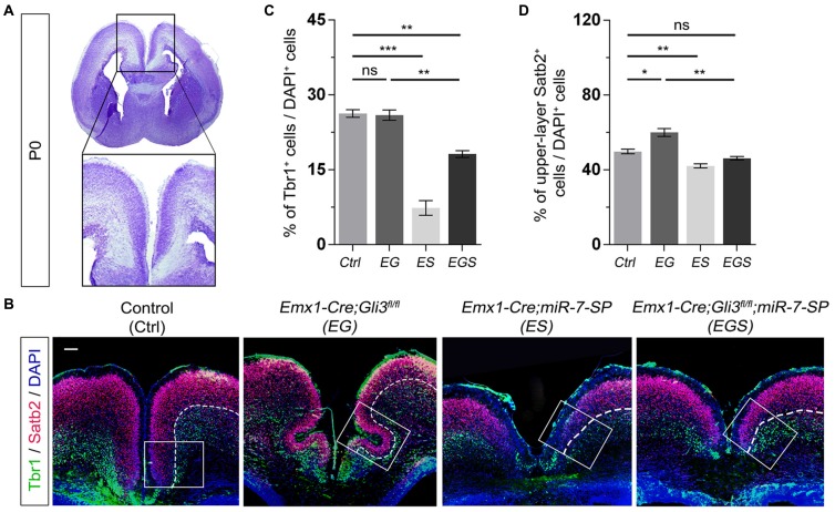
Figure 4.
Gli3 and miR-7 regulate population of newborn neurons in the cortical midline through an opposite effect. (A) The position that was captured and presented in the P0 cortical midline region. (B,C) Absence of Gli3 showed no alteration of Tbr1+/DAPI+ neurons in the deep layer. But losing function of both miR-7 and Gli3 significantly reduced the population of Tbr1+/DAPI+ cells. Knockdown of miR-7 significantly reduced the proportion of Tbr1+/DAPI+ neurons in the deep layer. (B,D) The upper layer neurons were separated according to the zones of Satb2+ intensive cell layer and Tbr1+ marked layer using the white dotted line. Cortical deficiency of Gli3 increased the proportion of upper layer Satb2+/DAPI+ cells, which was rescued by silencing miR-7. Knockdown of miR-7 significantly reduced the proportion of Satb2+/DAPI+ neurons in the upper-layer. The markers Tbr1, Satb2 and DAPI stained for newborn neurons in deeper layer, newborn neurons in upper layer and all cells, respectively. Values represent mean ± SEM. n > 9. *P < 0.05; **P < 0.01; ***P < 0.001; ns, not significant. One-way ANOVA with post hoc test was used. Scale bar = 100 μm.

