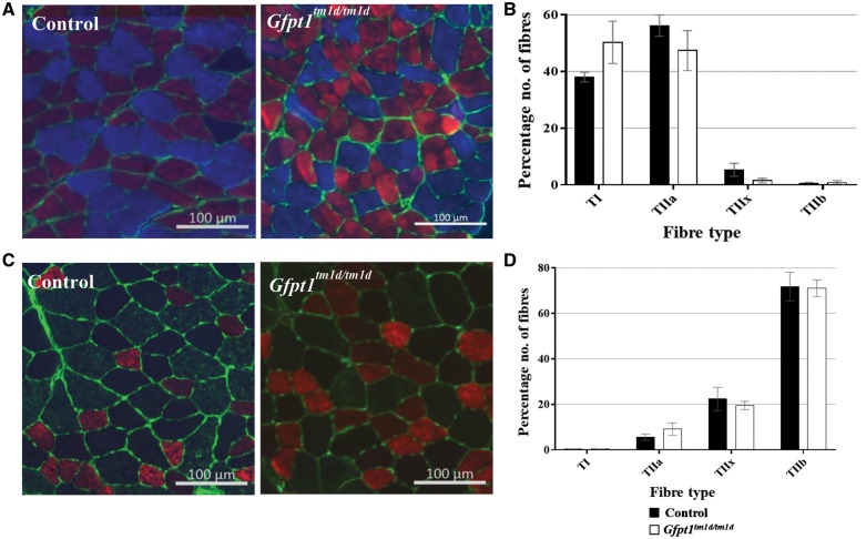
Figure 4.
Fibre type labelling in control and Gfpt1tmld/tmld mouse muscle. Soleus (A) and TA (C) muscles were labelled with antibodies against MHC: Type 1 (blue) I, Type IIa (red), Type IIb (green) and laminin (green). Type IIx fibres were labelled on a second section (not shown). There were no differences in the fibre type proportion in both the soleus (n = 4) (B) and TA muscles (n = 4) (D).

