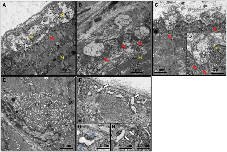
Figure 6.
Altered morphology at the ultrastructural level in Gfpt1tm1d/tm1d mouse muscle. Representative electron micrographs from 3 months old control and Gfpt1tm1d/tm1d intercostal muscles. Examples of control (A), and Gfpt1tm1d/tm1d(B–D) NMJs. Tubular aggregates (E) and subsarcolemmal vesicular structures (F–H) in Gfpt1tm1d/tm1d mouse muscle. Synaptic vesicles (*), junctional folds (red arrow), mitochondria (M), tubular aggregates (TA), subsarcolemmal vesicular structures (white arrow), dense filamentous-like material (blue arrowhead) (n = 4).

