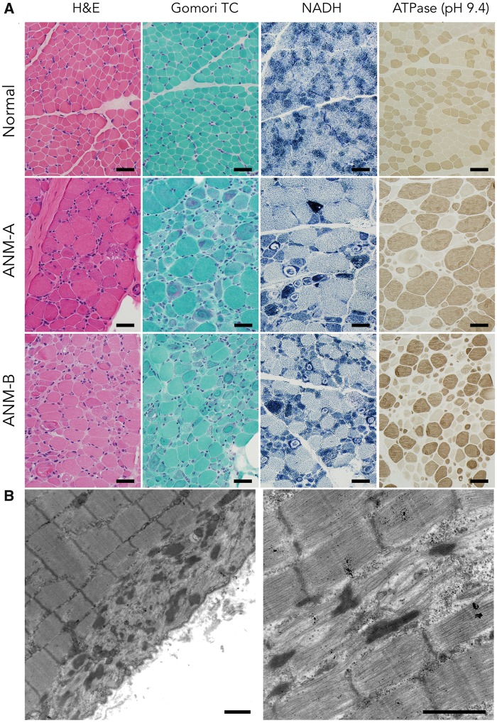
Figure 3.
Histological Findings in Human ANM. (A) In comparison to a normal muscle biopsy from the m. vastus lateralis muscle of a 17-month-old boy (upper panels), muscle biopsies from two ANM patients (ANM-A, middle panels; and ANM-B, lower panels) display marked abnormalities of myofiber size (both hypotrophy and hypertrophy) and increased numbers of internally nucleated fibers on hematoxylin and eosin (H&E) staining. Some fibers show areas of vacuolization. Gomori trichrome stain highlights aggregates of granular reddish-purple inclusions (nemaline rods), along with ring-like subsarcolemmal areas of myofibrillar disorganization. NADH stain (which highlights mitochondria and elements of the sarcotubular system) shows dense aggregates of these organelles in some fibers as well as areas of clearance within some of the ring-like subsarcolemmal inclusions. An ATPase stain at pH 9.4 (which causes fast type 2 fibers to stain dark brown and slow type 1 fibers to stain light tan) reveals a hypertrophic fiber population exclusively composed of Type 2 fast fibers amid a hypotrophic population of both fiber types (Bar=40 µm). (B) Ultrastructural appearance of nemaline rods (left panel) and myofibrillar disarray (right panel) in ANM muscle (Bar=1 µm).

