Figure 5.
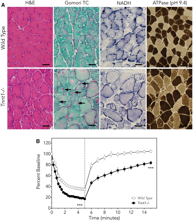
Muscle phenotype of Tnnt1−/− mice. (A) Compared with control mice (upper panels), quadratus femoris muscles from Tnnt1−/− (lower panels) mice have myofiber hypertrophy and hypotrophy and internal nucleation similar to that seen in humans with ANM (H&E). Gomori trichrome staining highlights the characteristic granular red nemaline rod aggregates (arrows). In contrast to findings seen in ANM patients, NADH stain shows appropriate distribution of organelles in Tnnt1−/− muscle. On ATPase staining (pH 9.4), large and small fiber populations include both slow and fast myofibers, another finding distinct from human ANM muscle (Bar=40 µm), in which hypertrophic fibers are exclusively Type 2. (B) Ex vivo functional measurement of whole soleus muscles demonstrates larger and faster loss of force during intermittent muscle fatigue contractions in Tnnt1−/− (solid circles) relative to wild-type (open circles) mice. When fatigue contractions terminate (dashed line), Tnnt1−/− muscles show slower and less complete recovery from fatigue (Tnnt1−/−, n = 3; WT, n = 5; values as mean±SE, comparison of means, ***P < 0.001 by two-way ANOVA with Bonferroni correction).

