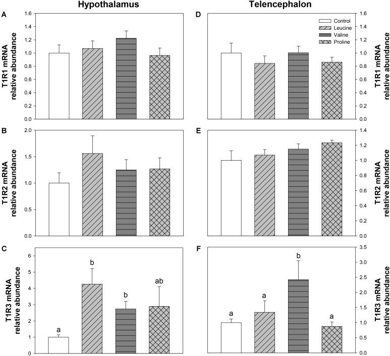
FIGURE 10.
Expression of T1R family taste receptors. mRNA levels of T1R1 (A,D), T1R2 (B,E), and T1R3 (C,F) in hypothalamus (A–C) and telencephalon (D–F) of rainbow trout 6 h after intraperitoneal administration of 0.5 mL⋅100 g-1 body mass of saline solution alone (control) or containing 40 μmol⋅mL-1 of L-leucine, 40 μmol⋅mL-1 of L-valine, or 40 μmol⋅mL-1 of L-proline. Each value is the mean + SEM of n = 6 fish per treatment. Gene expression results are referred to control group and are normalized by EF1α and β-actin expression. Different letters indicate significant differences (P < 0.05) between different groups. P-values are: for (A), = 0.403; for (B), = 0.463; for (C), C vs L = 0.015, C vs V = 0.029, C vs P = 0.386, L vs V = 0.202, L vs P = 0.414, V vs P = 1; for (D), = 0.642; for (E), = 0.346; and for (F), C vs L = 0.508, C vs V = 0.037, C vs P = 0.813, L vs V = 0.043, L vs P = 0.571, V vs P = 0.035.

