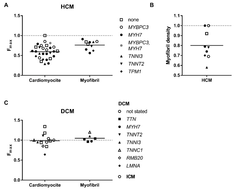
Figure 1.
Maximum force of contraction by different types of preparations: from hearts of patients with HCM (A) and DCM (C). As can be seen, the maximal force generated by cardiomyocytes as well as myofibrils is lower in the HCM samples. The force is not diminished in DCM samples. (B) Density of myofibrils in cells were measured in some HCM samples. Each data point represents a different experimental group where the symbols indicate genes where mutations were found. See also Table 1 and Table 2. All values are normalised to those of donor heart muscle (dashed line).

