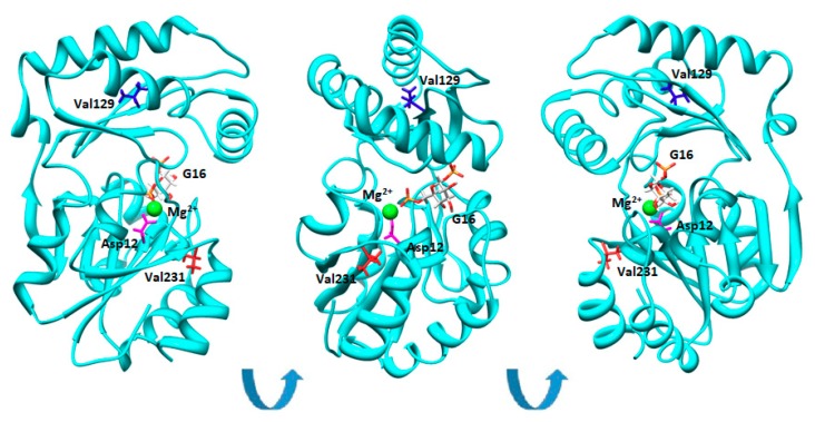
Figure 1.
Val129 and Val231 in the structure of phosphomannomutase2. Phosphomannomutase 2 (PMM2) (single chain) is represented as cartoons. Asp12, Val129, and Val231 are shown as sticks in magenta, blue, or red, respectively. Glucose-1,6-bisphosphate (G16) is represented by sticks and is colored by atom types. Mg2+ is represented by a green sphere. Different orientations of the same molecule are shown.

