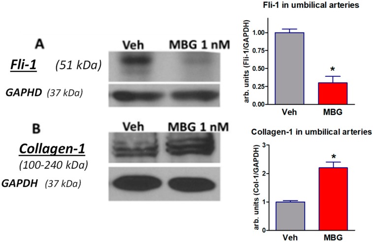
Figure 2.
Western blots of Fli-1 (A) and collagen-1 (B) in placentae from normotensive pregnant subjects incubated with vehicle (grey columns) or 1 nM MBG (red columns). Left—representative Western blots, right—bars representing means ± SEM from 4 densitometry measurements. By t-test: * p < 0.01 vs. vehicle. Levels of Fli-1 and of collagen-1 were normalized against levels of GAPDH.

