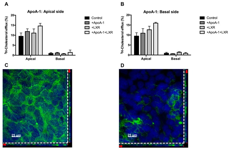
Figure 5.
Transport activity and localization of ABCA1 in human primary trophoblast cells polarized on Transwell® system. (A,B) The confluent trophoblast monolayer was loaded with 1 µCi of 3H-cholesterol at the apical side for 24 h. After equilibration, 10 µg/ml of apoA-1 was added to the apical panel (A) or the basal side panel (B) for 8 h. Data are shown as mean ± SD from a representative experiment. Details of the efflux procedure are described in Materials and Methods, and (C,D) representative immunofluorescence staining of ABCA1 in the confluent STB monolayer. The orthogonal projections through the Z-stack suggested that ABCA1 (green) is predominantly expressed at the apical side (Figure 5C) while it was barely detectable at the basal side (Figure 5D) of the STB monolayer. Nuclei were counterstained with 4’,6-diamidino-2-phenylindole (DAPI, blue). The horizontal and vertical red arrows on the orthogonal plane show the level of the selected stack. The upper level of the stack shown by the red label on the right corner of the image (Figure 5C) corresponds to the apical outer membrane. The bottom level stack marked by the red label on the right corner of the image (Figure 5D) corresponds to the basal outer membrane of the STB. Details of the staining procedure are described in Materials and Methods.

