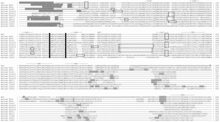
Figure 1.
Protein sequence alignment of A. pisum cysLGIC subunits. D. melanogaster RDLbd (RDL) is included for comparison. N-terminal signal leader peptides are shown in gray shading and white text. Loops implicated in ligand binding (LpA–F) are indicated, as well as the four transmembrane (TM) domains. The two cysteines forming the cys-loop are highlighted in black shading, and putative N-glycosylation sites are boxed. Potential cAMP, PKC, CK2 and tyrosine kinase phosphorylation sites are shown in gray shading. The sequences presented in this alignment can be found in the Supplementary Material.

