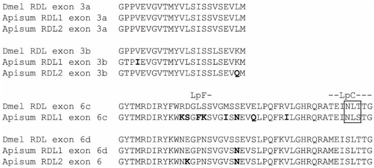
Figure 3.
Splice variants of A. pisum and D. melanogaster RDL. Alternative splicing of exons 3 and 6. Acyrthosiphon residues that differ from those of the orthologous Drosophila exon are highlighted in bold. N-glycosylation sites are boxed and Loops C and F, which contribute to ligand binding, are indicated.

