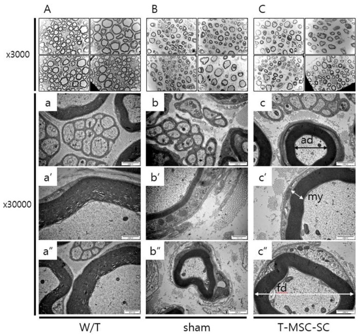
Figure 4.
Assessment of ultrastructure of sciatic nerves in Tr-J mice following transplantation with T-MSC-SCs by electron microscopy (EM). (A–C): ×3000 magnification; (a–c’’): ×30,000 magnification. Relative to wild-type (W/T) sciatic nerve, the myelinated nerve axon and large diameter axon were thinner in Tr-J mice than those in the sham and T-MSC-SC groups. ad (axonal diameter); fd (fiber diameter); my (myelin thickness).

