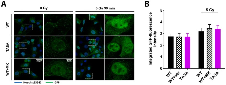
Figure 3.
The basal Akt phosphorylation is not required for its nuclear localization. TrC1 Akt1-TASA, Akt1-WT and Akt1-WT expressing cells pretreated with solvent or 4 µM MK-2206 (2 h before IR) were exposed to IR with 0 Gy or 5 Gy. 30 min after the irradiation cells were fixed in 3% paraformaldehyde, permeabilized with 0.2% Triton X-100 in phosphate-buffered saline (PBS), and subjected to immunofluorescence analysis. (A) Representative photomicrographs showing subcellular localization of the eGFP-coupled Akt1 variants Akt1-TASA and Akt1-WT upon the indicated treatments. The overview pictures were done using 63-fold magnification. Detailed pictures were obtained using 63-fold magnification. (B) Quantification of the integrated eGFP-intensity in nuclei of TrC1 expressing Akt1-TASA and Akt1-WT mutants with and without pre-treatment with the MK-2206 inhibitor. Quantification involves the analysis of 50 cells per condition and was performed by a CellProfiler software [19]. Data show means ± SD from 3 independent experiments; ANOVA test with Tukey correction and showed no significant differences.

