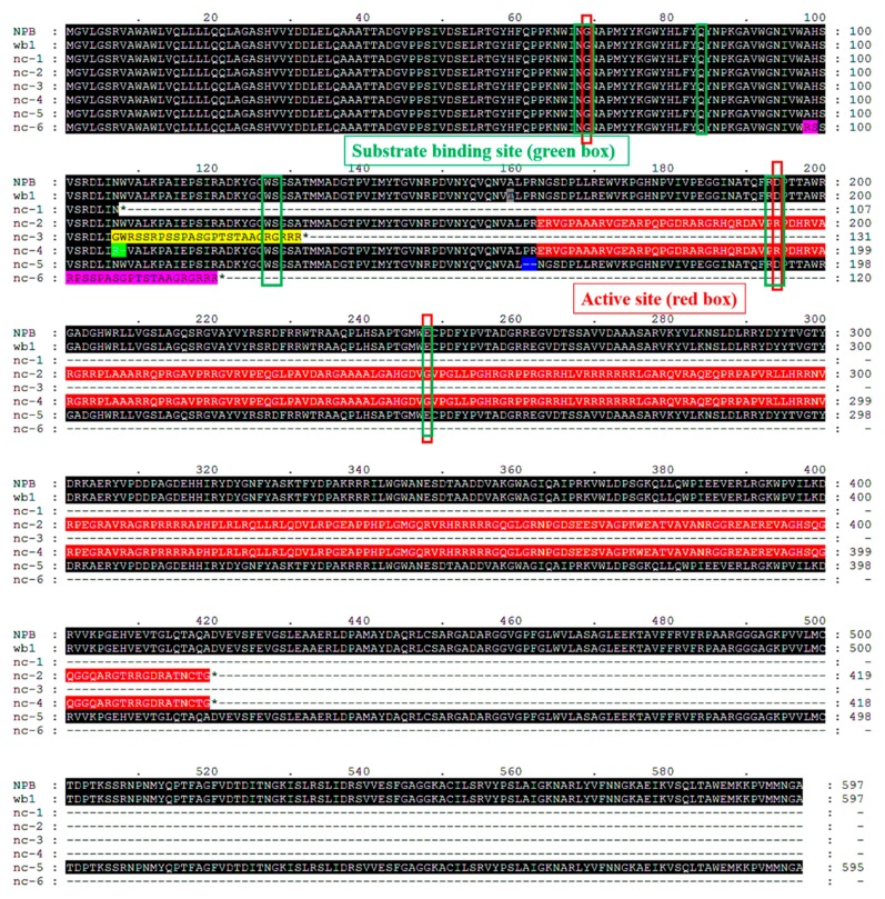
Figure 7.
WB1 alignments of NPB (WT) with six mutant lines and wb1. Analysis performed with MEGA 5.0 software. The substrate binding site (eight residues, green boxes) and the active site (three residues, red boxes) are indicated by the Blast search program (http://www.ncbi.nlm.nih.gov/BLAST/). Black color indicates a sequence that is consistent with that of WT. Except for the black color, the same color represents the same sequence, and different colors represent the different mutations among the mutant lines.

