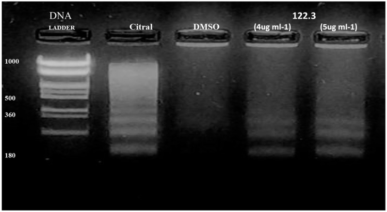
Figure 2.
Analysis of genomic DNA fragmentation in BS-24-1 cells. 2.5 × 10 5 cells were incubated for 24 h with different concentrations of fraction 122.3. Lane 1, 1KB ladder; Lane 2, positive control (5 μg/mL of citral for 1.5 h); Lane 3, negative control (DMSO); Lanes 4 and 5, cells treated with fraction 122.3 at the concentrations of 4 and 5 μg/mL, respectively.

