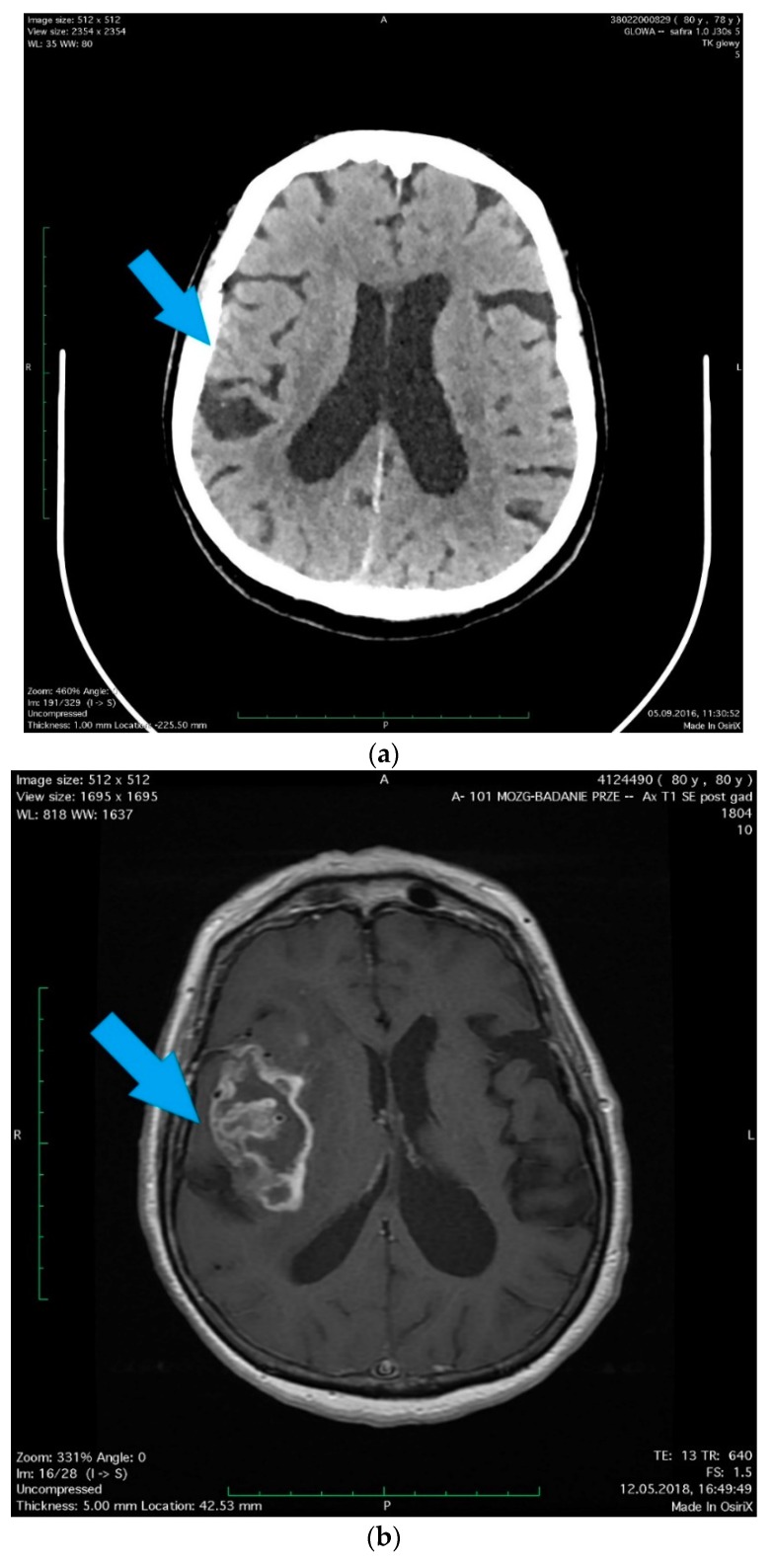
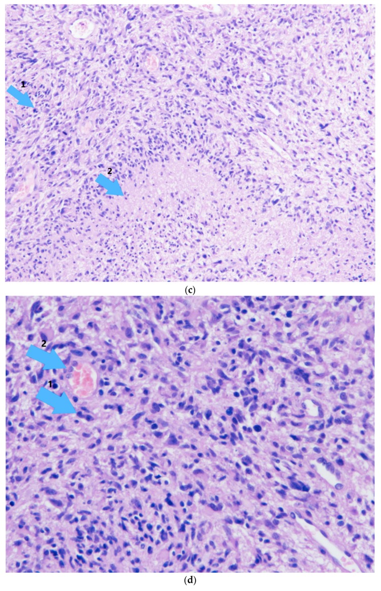
Figure 1.
A 78-year-old woman with hypertension and hypercholesterolemia was admitted to the hospital due to her head injury (after falling in moving bus), small changes were revealed in the computed tomography (CT) scan as well as in the radiographic examination that did not require surgical treatment. (a) (arrow) A little amount of blood along the falx of the brain, on the tent of the cerebellum on the right side and in a fissura of the right parietal lobe with scant concussion in it. In the white matter of corona radiata, a number of dimly defined, diminished-density areas were observed, most likely corresponding to degenerative vascular changes. The patient, without neurological deficits except diminishing post-traumatic headaches, was discharged after the observation period in a good condition. Two years later, the patient was readmitted to the Department of Neurosurgery with a diagnosed brain tumor, manifested by a 1.5-month history of memory and orientation disorders. (b) (arrow) In the MRI scans, the right brain hemisphere tumor was found, the position of which corresponded to previous post-traumatic lesions. Right temporoparietal craniotomy was performed and the cytoreduction of the tumor was extended. Histopathology revealed Glioblastoma multiforme (facultatively). The histopathological pictures show (c) characteristic necrosis, surrounded by a pseudopalisade (arrow 1), visible cell mitosis (arrow 2), (d) large cell density, mitosis (arrow 1), lumen of the vessel with erythrocytes (arrow 2). The patient was accepted for radiotherapy with chemotherapy and discharged home in good condition, without additional neurological deficits, walking with little assistance. The study was approved by the Local Ethical Committee at the Pomeranian Medical University in Szczecin, Poland (approval No. KB-0012/96/14; Szczecin, Poland, 24.11.2014).


