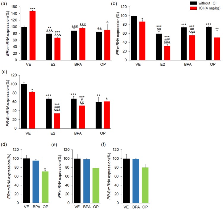
Figure 5.
E2, BPA, and OP effects and estrogen and progesterone receptor expressions in maternal uterus and implantation sites. The expression levels of ERα, PRα, and PRβ genes were measured by real-time PCR and normalized to that of 18S ribosomal RNA (RN18S). In uterus, (a) mRNA levels of ER were not changed by BPA or OP with/without ICI; (b) mRNA levels of PR were significantly low in all groups; (c) mRNA levels of PR-B were markedly low in all groups; In implantation sites, (d–f) the expressions of ERα mRNA were significantly decreased by OP. Statistical significance was determined by two-way ANOVA. * p < 0.05 vs. VE, ** p < 0.01 vs. VE, *** p < 0.001 vs. VE, # p < 0.05 EDs + ICI vs. EDs, ## p < 0.01 EDs + ICI vs. EDs, ### p < 0.001 EDs + ICI vs. EDs, & p < 0.05 vs. ICI, && p < 0.01 vs. ICI, &&& p < 0.001 vs. ICI. n = 5 mice per group for uterus, n = 3 mice per group for implantation sites. Treatments: E2; 40 µg/kg/day, BPA; 100 mg/kg, OP; 100 mg/kg, ICI; 4 mg/kg.

