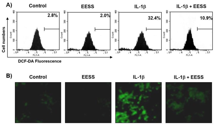
Figure 3.
Effects of EESS on IL-1β-induced ROS generation in rat articular chondrocytes. Cells were pretreated with 300 μg/mL EESS for 1 h, and then stimulated with or without 40 ng/mL IL-1β for 1 h. Cells were incubated with 10 μM DCF-DA for 30 min at 37 °C. (A) The cells were collected, and DCF fluorescence was measured by flow cytometry. Values represent the means of two independent experiments. (B) Images were obtained by fluorescence microscopy, and those presented are from one experiment, and are representative of at least three independent experiments.

