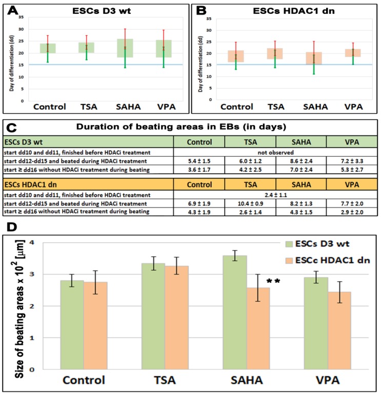
Figure 1.
The beating of cardiomyocytes in differentiated HDAC1 wild-type (wt) and HDAC1 double knockout (dn) mouse embryonic stem cells (ESCs). Colonies of ESCs (embryoid bodies, EBs) were monitored every day in (A) HDAC1 wt mESCs, and (B) HDAC1 dn mESCs. Bars in panels (A,B) show an average day of cardiomyocytes’ beating. Green standard errors (scales) show a start of beating and red standard errors (scales) show the end of beating (scale of the y-axis is in days). The observation was performed in non-treated wt and HDAC1 dn mESCs induced to cardiac differentiation and these cells treated by HDACi (TSA, SAHA, and VPA). (C) A duration of beating areas in EBs of non-treated and EBs treated by TSA, SAHA, and VPA is shown. (D) A size of beating areas in embryonic bodies is documented. Asterisks (**) show a statistically significant difference at p ≤ 0.05.

