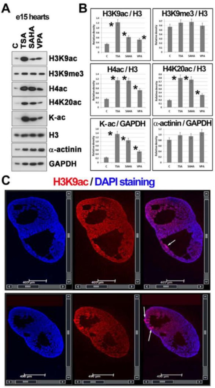
Figure 5.
Histone post-translational modifications studied in mouse embryonic hearts (e15) treated with HDACi. (A) Western blots showed changes in H3K9ac, H3K9me3, H4ac, H4K20ac, pan-acetylated lysines (K-ac), and α-actinin in e15 embryonic hearts treated with HDACi (TSA, SAHA, and VPA). Data on histone levels were normalized to the level of histone H3 and non-histone proteins were normalized to the level of GAPDH. An identical protein amount for each experimental event was loaded on the gel. (B) Data from panel (A) were normalized to the relevant reference protein GAPDH, and the density of Western blot fragments was statistically analyzed using Student’s t-test; asterisks show statistically significant differences at p ≤ 0.05. GAPDH was used for data normalization, and α-actinin was used as a marker of cardiomyocytes. (C) The distribution pattern of H3K9ac (red) in the e15 mouse embryonic hearts is shown. DAPI (blue) was used as a counterstain of the cell nuclei. Arrows show the accumulation of H3K9ac in ventricular portions.

