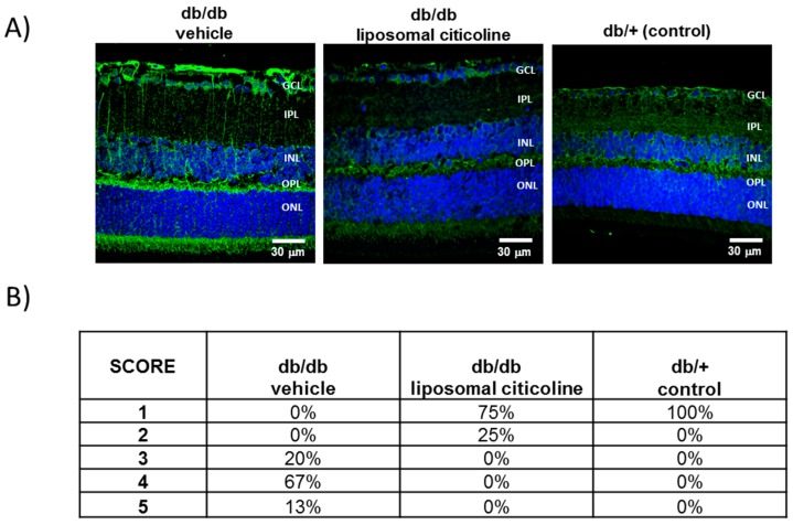
Figure 2.
Effect of citicoline on glial activation. (A) Comparison of GFAP immunoreactivity (green) in the retina among representative samples from a diabetic mouse treated with vehicle, a diabetic mouse treated with citicoline, and a non-diabetic mouse. Nuclei were labeled with Hoechst (blue). ONL: outer nuclear layer; OPL: outer plexifom layer; INL: inner nuclear layer; IPL: inner plexiform layer; GCL: ganglion cell layer. Scale bars, 30 µm. (B) Quantification of glial activation based on the extent of GFAP staining. n = seven mice per group.

