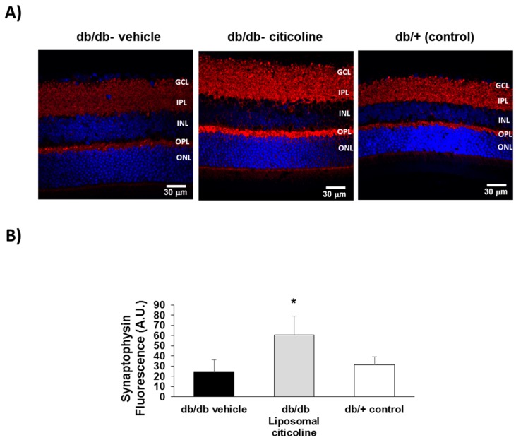
Figure 6.
Effect of citicoline on synaptophysin. (A) Comparison of synaptophysin immunofluorescence (red) between representative samples from a db/db mouse treated with vehicle, a db/db mouse treated with citicoline, and a non-diabetic mouse. Nuclei were labeled with Hoechst (blue). ONL: outer nuclear layer; OPL: outer plexiform layer; INL: inner nuclear layer; IPL: inner plexiform layer; GCL: ganglion cell layer; (B) Quantification of synaptophysin immunofluorescence in arbitrary units (AU). Results are mean ± SD. * p < 0.01 vs. the other groups.

