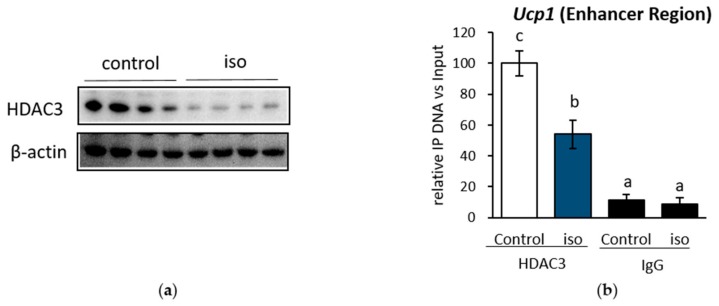
Figure 5.
HDAC3 protein level was decreased under β-AR stimulation in IWAT cell. (a) HDAC3 protein level, and its recruitment level in (b) the Ucp1 enhancer region after treatment with 10 μM iso for 4 h. Data are presented as mean ± S.E.M. (error bars). n = 4–6 in each group. β-actin act as loading control and IgG as a mock control. Different letters indicate significant differences (p < 0.05) according to one-way ANOVA followed by the Tukey-Kramer multiple comparison test. Same letters indicate non-significant difference.

