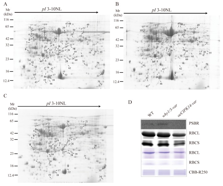
Figure 1.
2-DE and immunoblot analysis of total proteins extracted from rosette leaves of WT and two variegated mutants. (A–C) Representative 2-DE gel images of WT (A); why1/3-var (B); and oeCIPK14-var (C). An equal amount (1.5 mg) of total proteins was loaded on each IPG strips (3–10 NL). The spot numbers indicated proteins that showed significant changes between WT and two variegated mutants. (D) The changing of protein abundance selected from 2-DE were confirmed by western blot and CBB R250 staining. The immunoblot analysis is performed using antibodies against RBCL, RBCS, and PSBR. CBB R250 staining shows RBCL and RBCS protein amount and the same amount of loading proteins.

