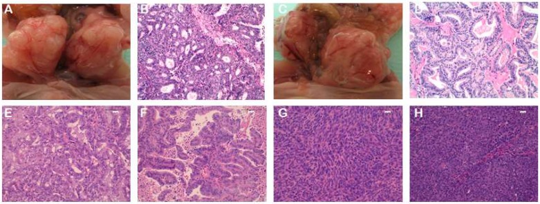
Figure 2.
Representative PDX images from orthotopic (A–D) and heterotopic (E–H) models. (A–D) Orthotopic PDX from two different endometrioid EC patients. Panels A and C are a macroscopic image of the tumour growth in the uterus. Panels B and D represent images of the H&E staining of the PDX tumours. (E,F) H&E stainings from subcutaneous PDX models of two different endometrioid EC patients. (G,H) H&E stainings from subcutaneous PDX models of two different non-endometrioid EC patients. Panel G corresponds to a carcinosarcoma histology, and panel H corresponds to a serous carcinoma. Magnification 20×.

