
Fig. 2.
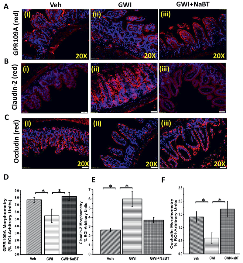
Change in gut microbiome in GWI alter niacin receptor (GPR109A) and tight junction proteins in the intestine. A. The expression pattern of butyrate and niacin receptor GPR109A was assessed by immunofluorescence microscopy. The representative images showed immunoreactivity of GPR109A in the distal part of the small intestine of veh control group of mice (veh, n = 3), gulf war illness group of mice (GWI, n = 3) and a group of mice co-exposed with GWI and sodium butyrate (GWI + NaBT, n = 3). B and C. The expression pattern of Claudin-2 and Occludin (tight junction proteins) was assessed by immunofluorescence microscopy. Tissue levels of Claudin-2 (B) and Occludin (C) in Vehicle control group of mice (Veh, n = 3), gulf war chemical treated group of mice (GWI, n = 3) and a group of mice co-exposed with GWI and sodium butyrate (GWI + NaBT, n = 3) was assessed by immunofluorescent microscopy after labeling the protein with the red fluorescent secondary antibody and counterstained with DAPI (blue). D–F. The bar diagram shows the quantitative morphometric analysis of fluorescence intensities of GPR109A (D), Claudin-2 (E), and Occludin (F) immunoreactivity in the region of interest (ROI) in the small intestine. *(p < 0.05). (For interpretation of the references to colour in this figure legend, the reader is referred to the web version of this article.)