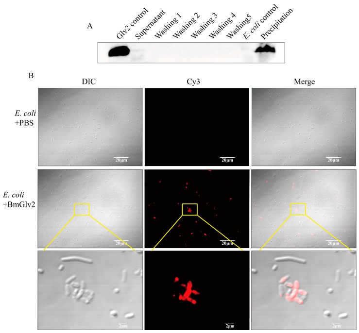
Figure 6.
The binding activity analysis of recombinant BmGlv2. (A) Western Blotting analysis of the binding of BmGlv2 to E. coli JM109 cells. Glv2 control, recombinant Glv2; Supernatant, a supernatant fraction after binding incubation; Washing1–Washing5, the first to fifth washing collection; E. coli control, E. coli JM109 only; Precipitation, the centrifuged E. coli cells after incubation and washing. The E. coli JM109 was incubated with BmGlv2 for 3 h. (B) Immunofluorescence localization analysis of BmGlv2 on the surface of E. coli JM109 cells. Confocal microscopy was performed to visualize BmGlv2 binding (red) on the cell walls of E. coli JM109. The third row amplified from the second row described the detail of BmGlv2 on the surface of E. coli JM109 cells. The PBS was used as a negative control. The scale bar was marked in the picture.

