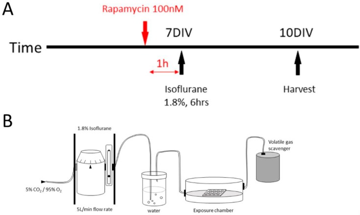
Figure 2.
Schematic representation of the experimental timeline and exposure induction diagram in vitro. (A) The general experiment timeline in vitro. The neurons were exposed to 1.8% isoflurane for 6 h on their 7 DIV, and 100 nM rapamycin was added into the media 1 h before the exposure according to the experiment design. The fresh media change was done regularly. The cells were fixed for immunohistochemistry on 10 DIV; (B) Coverslips in 12-well plates were placed in identical air-tight, humidified chambers. Isoflurane was delivered using an agent-specific, calibrated inline and was diluted in 5% CO2/95% O2 carrier gas. Controls for these experiments received 5% CO2/95% O2 carrier gas only. After a 15-min equilibration period, the sealed chambers were placed in an incubator to maintain a temperature at 37 °C for the duration of the anesthesia exposure.

