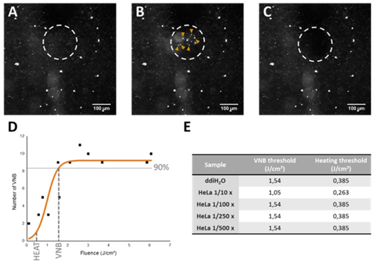
Figure 5.
Determination of heating and VNB threshold via dark-field microscopy. (A) dark-field microscopy image of JetPEI/pDNA/AuNP 5 pt complexes in ddiH2O; (B) dark-field microscopy image upon VNB formation (VNBs indicated by yellow arrows); (C) dark-field microscopy image after VNB formation. Scalebar on the images represents 100 μm; (D) graph shows the relation between the number of VNBs and laser fluence. VNB threshold is calculated as the laser fluence needed to reach 90% of the maximum number of VNBs. The fluence for heating is selected at one-fourth of the VNB threshold; (E) the table shows the threshold values for VNB formation and heating (in J/cm2) used for JetPEI/pDNA/AuNP 5 pt complexes in further experiments.

