Fig. 8.
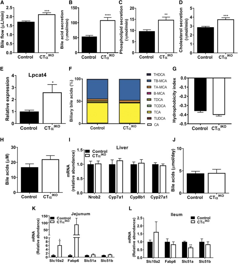
Loss of intestinal CTα enhances biliary bile acid secretion. Gallbladders of male control and CTαIKO mice were cannulated, and bile was collected in preweighed containers (n = 6–8 per group). A: Bile flow. B: Bile salt secretion. C: Phospholipid secretion. D: Cholesterol secretion. E: Jejunal Lpcat4 expression. F: Relative biliary bile acid composition. G: Bile acid hydrophobicity index. H: Plasma bile acids. I: mRNA abundance of hepatic genes involved in bile acid synthesis. J: Fecal bile acids. mRNA abundance of jejunal bile-acid responsive genes (K) (n ≥ 4 per group) and mRNA abundance of ileal bile acid-responsive genes in control and CTαIKO mice 2 h after refeeding the HFD (L) (n = 3 per group). Values are means ± SEM. * P < 0.05; ** P < 0.01; *** P < 0.001; **** P < 0.0001. Β-MCA, β-β-muricholic acid; CA, cholic acid; Cyp7a1, cytochrome P450, family 7 subfamily a member 1; Cyp8b1, cytochrome P450, family 8 subfamily b member 1; Cyp27a1, cytochrome P450, family 27 subfamily a member 1; Fabp6, FA binding protein 6; Nrob2, nuclear receptor subfamily 0 group b member 2; TA-MCA, tauro-α-muricholic acid; TB-MCA, tauro-β-muricholic acid; TCA, taurocholic acid; TCDCA, taurochenodeoxycholic acid; THDCA, taurohyodeoxycholic acid; TUDCA, tauroursodeoxycholic acid.

