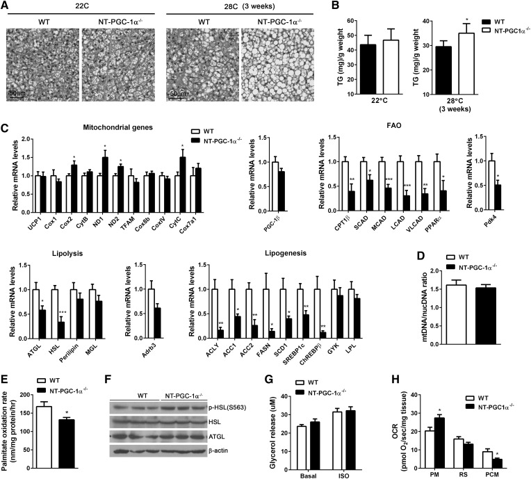
Fig. 2.
FAO is lower in BAT of NT-PGC-1α−/− mice housed at thermoneutrality. A: Representative H&E images of BAT from 7 week old WT and NT-PGC-1α−/− mice housed at 22°C (22C) or 28°C (28C) for 3 weeks (n = 4 or 5 per group). Scale bars, 50 µm. B: Triacyglycerol (TG) content of BAT from WT and NT-PGC-1α−/− mice (n = 8 per genotype). C: qPCR analysis of BAT gene expression in WT and NT-PGC-1α−/− mice housed at 28°C for 3 weeks (n = 8 per genotype). D: qPCR analysis of mitochondrial biogenesis (28°C). The relative ratio of mitochondrial DNA (mtDNA) to nuclear DNA (nucDNA) was analyzed (n = 8 per genotype). E: The [14C] palmitate oxidation rates measured in fresh BAT homogenates in triplicate (n = 7 per genotype) (28°C). F: Western blot analysis of lipolytic enzymes in BAT. G: Basal and isoproterenol-stimulated lipolysis of BAT tissues (28°C). Glycerol release was measured in the absence and presence of isoproterenol (ISO; 1 µM) (n = 7 or 8 per genotype). H: OCRs of BAT explants in the presence of malate/pyruvate (PM), rotenone/succinate (RS), or malate/palmitoyl carnitine (PCM) (28°C). Measurements were performed in duplicate (n = 5 per genotype). All data are presented as mean ± SEM. *P < 0.05; **P < 0.01; ***P < 0.001 determined by Student’s t-test.

