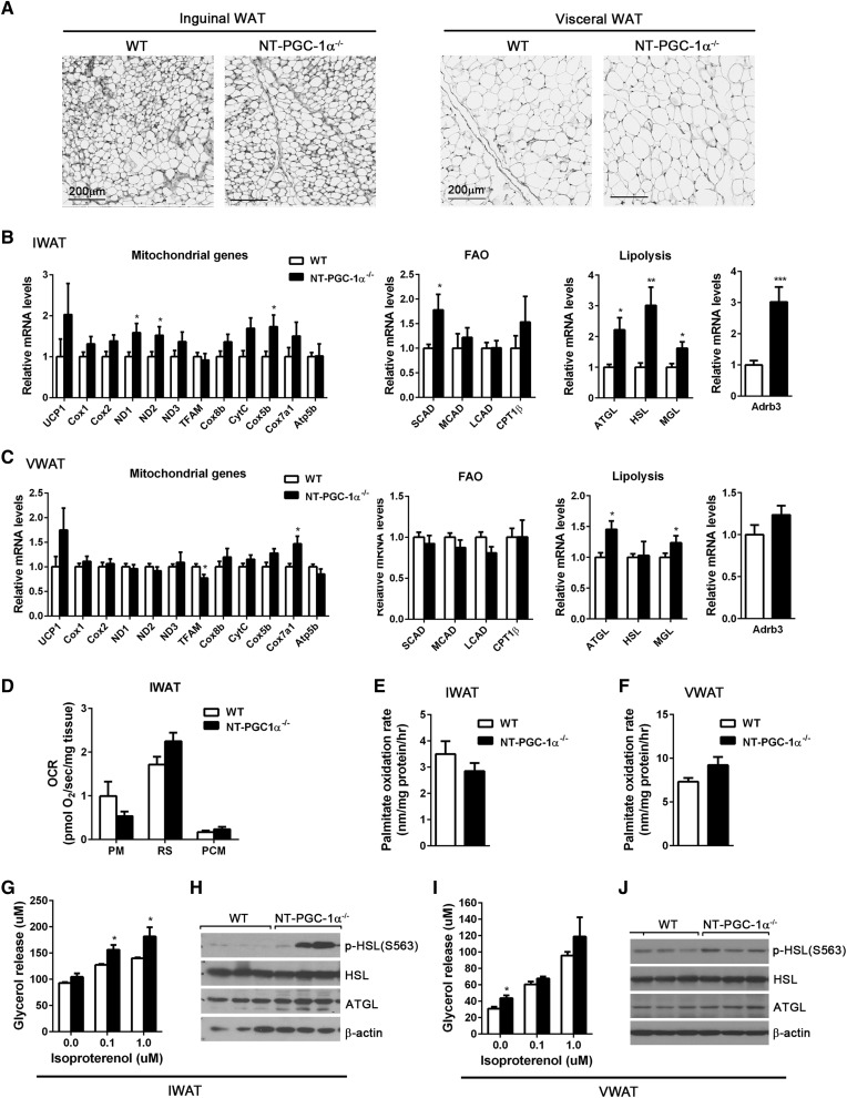
Fig. 3.
Effects of NT-PGC-1α ablation on WAT. A: Representative H&E images of inguinal and visceral (gonadal) WAT from WT and NT-PGC-1α−/− mice adapted at 28°C for 3 weeks. Scale bars, 200 µm. B, C: qPCR analysis of gene expression in IWAT and visceral WAT (VWAT) from WT and NT-PGC-1α−/− mice (n = 8 per genotype). D: OCRs of IWAT explants in the presence of malate/pyruvate (PM), rotenone/succinate (RS), or malate/palmitoyl carnitine (PCM). Measurements were performed in duplicate (n = 5 per genotype). E, F: [14C] palmitate oxidation rates of fresh IWAT and VWAT homogenates (n = 7 per genotype). G, I: Basal and isoproterenol-stimulated lipolysis of primary adipocytes isolated from fresh IWAT and VWAT. Glycerol release was measured in the absence and presence of isoproterenol (0.1 and 1 µM) (n = 7 or 8 per genotype). H, J: Western blot analysis of lipolytic enzymes in IWAT and VWAT. All data are presented as mean ± SEM. *P < 0.05; **P < 0.01; ***P < 0.001 determined by Student’s t-test.

