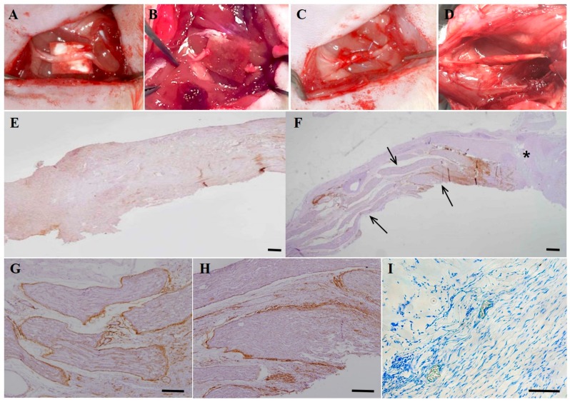
Figure 6.
Schematic images and immunohistochemical analysis of in vivo cell transplantation in experimental rats with nerve defects. (A) The sciatic nerve was exposed on the thigh of the experimental animal; (B) The sciatic nerve was resected and a 5-mm nerve gap developed; (C) After tubulization with biodegradable collagen bovine dura mater (Lyoplant®) in the nerve gap, a total of 1 × 10⁶ cells (DF-chN) were transplanted into the collagen tube with a 0.1-mL fibrin glue scaffold (Greenplast®). The tube was then sealed with sutures; (D) Photograph of the experimental nerve at eight weeks after DF-chN transplantation. In the cell transplantation site, the regeneration of nerve defect was detected with continuity of the nerve gap; (E–I) Histological and immunohistochemical analysis of low-affinity nerve growth factor receptor (p75NGFR) in regenerated motor nerve fibers at eight weeks post cell transplantation. (E) In non-cell-transplanted control specimens, regenerated nerve fibers were not detected in abundance and p75NGFR expression was weak. (F) In the DF-chN transplanted group, significantly increased regenerating nerve fibers were detected in the nerve gap (arrows). The asterisk (*) indicates the proximal end of the nerve gap. (G,H) High magnification of generated nerve fibers at the DF-chN graft site. High expression of p75NGFR was detected in newly generated nerve fibers, especially in the perineurium and endoneurium of regenerating nerve fibers. (I) Toluidine blue staining of regenerating nerve fibers showed blue-colored positive staining of the myelin sheath, which was detected in abundance in the outer layer of newly generated axonal fibers (Scale bar = 100 µm).

