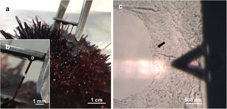
Figure 1.
a) Collection of Paracentrotus lividus footprints on mica. b) Detailed view of a sea urchin tube foot attached to mica, showing the adhesive disc (D) and the stem (S). c) Optical microscopy (10×) illustrating the positioning of the moist adhesive footprint (indicated by the arrow) beneath the triangular-shaped AFM cantilever.

