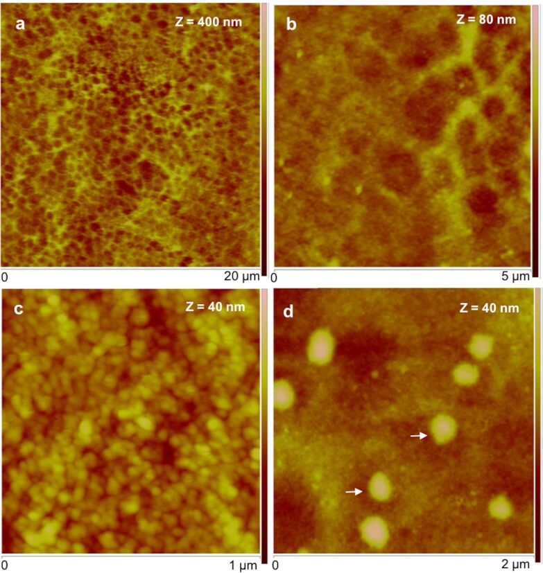
Figure 3.
Peak force tapping AFM (PFT-AFM) images of moist adhesive material deposited by the tube feet of Paracentrotus lividus on mica. a) Height image observed in air. b) Higher resolution of the same area showing the honeycomb appearance of the meshwork. c) Detailed topography view showing that the meshwork is composed of aggregated globular nanostructures. d) Detailed topography view of a different area showing bigger globular structures denoted by white arrows. Images were obtained with a ScanAsyst-Air probe.

