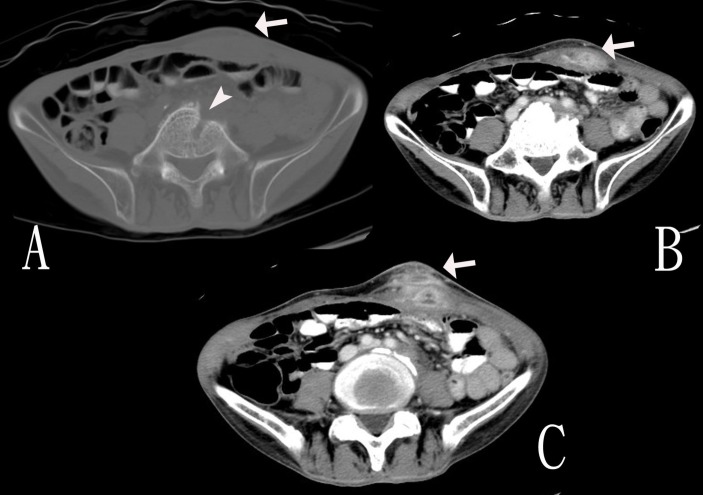
Figure 1.
Images of the third set of CT of the abdomen. (A) Bone window image shows a lytic tract at L5 vertebral body (arrow head). (B) Contrast-enhanced view shows the left lower abdominal wall abscess (arrow). (C) Contrast-enhanced view shows the left lower abdominal wall abscess with intraperitoneal extension (arrow).

