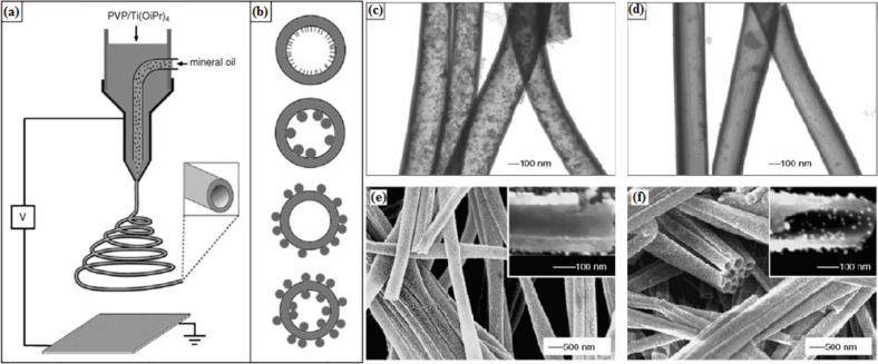
Figure 5.
(a) Schematic illustration of the setup using a dual-capillary spinneret to directly electrospin hollow nanofibers with functionalized surfaces; (b) schematic drawings showing cross sections of hollow nanofibers whose surfaces are derivatized with functional molecules (the top plate) and nanoparticles (NPs) (the other plates). TEM images of hollow titania nanofibers immersed in an oil-based ferrofluid overnight. The hollow fibers are prepared by co-electrospinning, either with (c) or without (d) octadecyltrichlorosilane (OTS) added to the mineral oil. (e) SEM image of hollow fibers for which inner and outer surfaces are derivatized with CH3- and NH2-terminated silanes, respectively, and then immersed in citrate-stabilized Au colloids. Note that the Au colloids are selectively adsorbed onto the outer surfaces. (f) SEM image of hollow fibers with inner and outer surfaces treated with an NH2-terminated silane, followed by incubation with Au colloids. In this case, the Au colloids are adsorbed on both surfaces. Reproduced with permission from [73], copyright 2005 Wiley.

