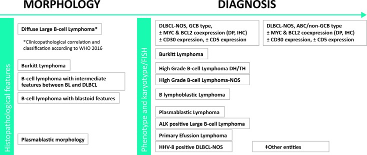
Figure 1. Histopathological diagnosis of DLBCL.
Tissue biopsy. (‡) Other entities: [T cell/histiocyte-rich large B-cell lymphoma, primary CNS large B-cell lymphoma, primary “leg type” large B-cell lymphoma, EBV-positive large B-cell lymphoma-NOS, DLBCL associated with chronic inflammation, lymphomatoid granulomatosis, primary mediastinal large B-cell lymphoma (thymic), Intravascular large B-cell lymphoma, Burkitt-like lymphoma with 11q aberration, B-cell lymphoma, Unclassifiable, with features intermediate between Hodgkin´s lymphoma and DLBCL (grey zone B-cell lymphoma), large B-cell lymphoma with IRF4 rearrangement]. DLBCL: large B-cell lymphoma; NOS: Not otherwise specified.

