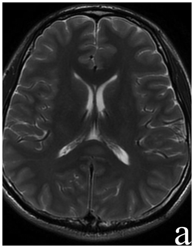
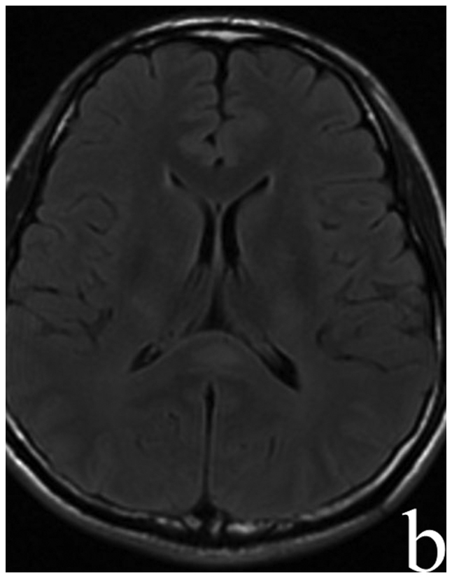
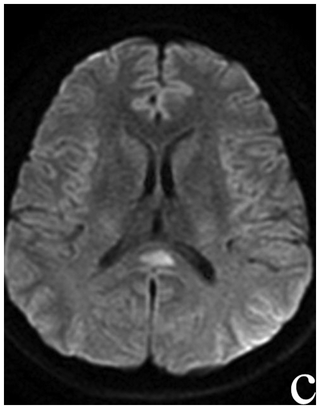
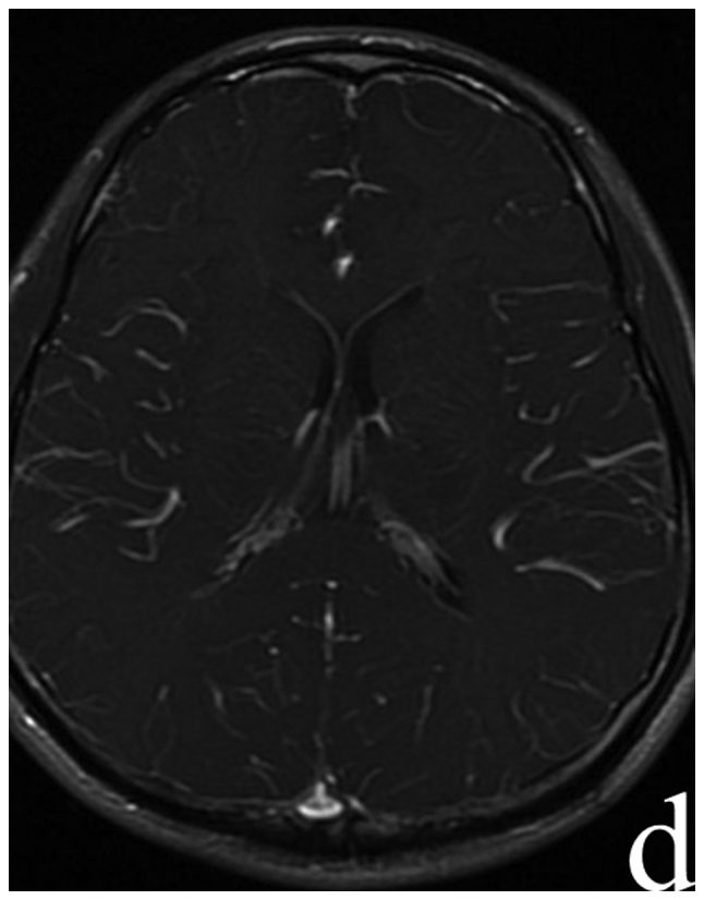
Figure 5.


Patient 3 MRI: (A) T2W, (B) FLAIR, (C) DWI sequences showed hyperintensity signal in the splenium of corpus callosum and (D) T1W enhanced scanning showed no enhancement effect. MRI, magnetic resonance imaging; FLAIR, fluid attenuated inversion recovery; DWI, diffusion-weighted imaging.
