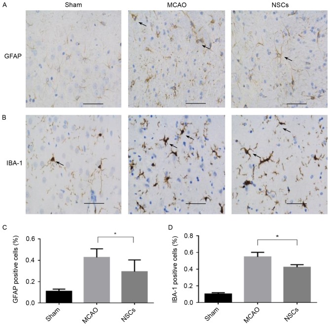
Figure 5.
The effect of NSC treatment on immunoreactive cells from the brain tissue of rats that underwent MCAO. (A) Immunofluorescence staining of GFAP. Arrows indicate GFAP positive cells. (B) Immunofluorescence staining of IBA-1. Arrows indicate IBA-1 positive cells. (C) Percentage of GFAP positive cells. (D) Percentage of IBA-1 positive cells. Data are presented as the mean ± standard deviation (n=5). *P<0.05. Scale bar=50 µm. Magnification, ×200. MCAO, middle cerebral artery occlusion group; NSC, neural stem cell group; GFAP, anti-glial fibrillary acidic protein; IBA-1, anti-ionized calcium binding adaptor protein.

