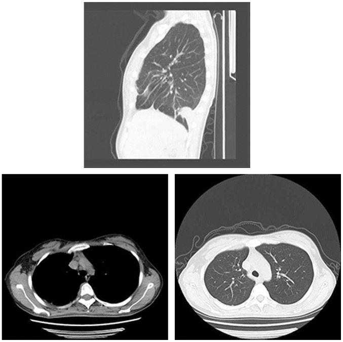
Figures 1.
CT images of patients with tuberculous pleurisy. Thorax is symmetrical and mediastinum is in the middle. Lung texture is natural. The right lung is scattered in a strip-like high-density shadow. The right side of the pleura shows nodular thickening. Trachea and bronchi are unobstructed, with no thickening of the wall of the tube. There were no enlarged lymph nodes and abnormal lumps in the mediastinum. The size and shape of the heart were normal, and no abnormal changes were seen in the vasculature. Right lung shows a patchy shadow, possibly indicating right pneumonic lesion and right pleural effusion. Right pleural nodules were thickened, indicating tuberculous pleurisy or pleural lesions. CT, computed tomography.

