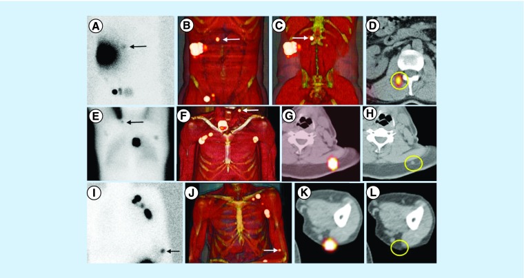
Figure 3. . Sentinel nodes in unexpected areas of lymphatic drainage.
(A) Anterior planar image of a patient with a melanoma in the right flank showing not only drainage to the right groin but also to the medial area of the trunk (arrow). Note that the sentinel node is clearly defined on (B & C) 3D imaging and (D) transversal SPECT/CT in the paravertebral muscle on the right. (E) In another patient with a melanoma of the back, posterior planar imaging shows drainage to both axillae and the left extracurricular region (arrow). This sentinel node is anatomically indicated on (F) 3D imaging and transversal (G) SPECT/CT and corresponds on (H) CT with a slightly enlarged node (circle), which contained metastases at histopathology. (I) In another patient with a melanoma of the left underarm, anterior planar imaging shows drainage to the left axilla and to the area of the left elbow (arrow). (J–L) On SPECT/CT, this sentinel node is seen as a dorsal subcutaneous node.
SPECT/CT: Single-photon emission computed tomography images with integrated computed tomography.
Reproduced with permission from [1] © Future Medicine Ltd. (2014).

