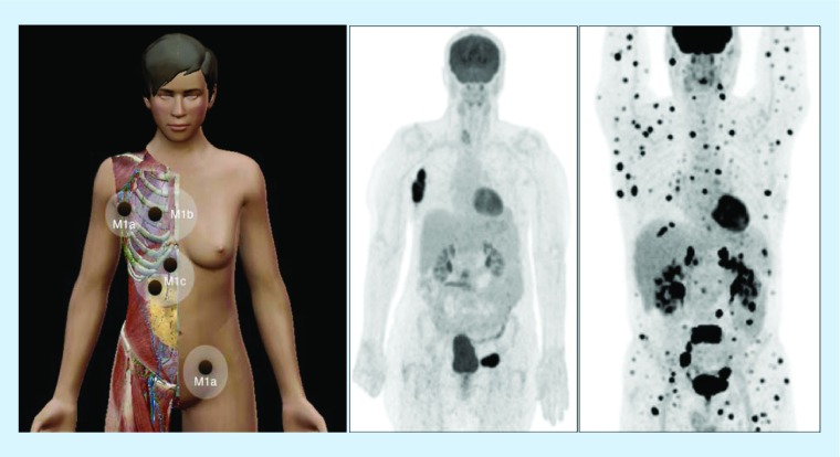
Figure 7. . Melanoma dissemination.
On the left, scheme illustrating disseminated disease in melanoma with distant metastases in lymph nodes (M1a), lung (M1b) and liver/bowel (M1c). Middle, 18F-FDG-avid lymph node metastases in right axilla and left groin as displayed on PET with maximum intensity projection. On the right, multiple FDG-metastases in another patient with widespread disease.
FDG: 18F-fluorodeoxyglucose.

