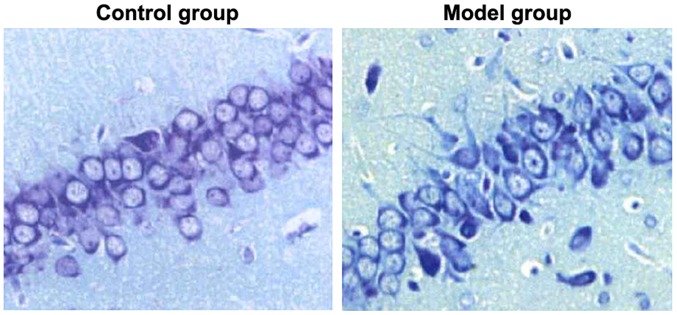
Figure 2.
Injuries of hippocampal neurons of rats in two groups detected through Nissal staining. Blue refers to nuclei. The hippocampal neurons of rats in control group are compactly arranged, with complete cell morphology; the hippocampal neurons of rats in model group are loosely arranged, with fuzzy cell outline.

