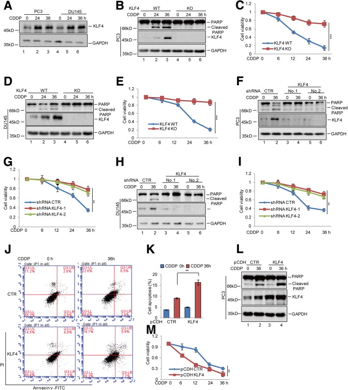
Fig. 1.
KLF4 enhanced cisplatin-induced apoptosis in prostate cancer cells. a PC3 and DU145 cells were treated with 20 μM cisplatin (CDDP) at the indicated times. The protein levels of KLF4 were analysed by western blotting. b-e PC3 and DU145 cells with or without KLF4 knockout (KO) were treated with 20 μM cisplatin at the indicated times. Cell apoptosis was detected by western blotting. Cell viability was measured by a CCK8 assay. Data represent the mean ± SD of three independent experiments. ***p < 0.001 vs. control. f-i PC3 and DU145 cells with or without KLF4 knockdown were treated with 20 μM cisplatin at the indicated times. Cell apoptosis was detected by western blotting and cell viability was measured by a CCK8 assay. Data represent the mean ± SD of three independent experiments. **p < 0.01 vs. control. j-m PC3 cells with or without KLF4 overexpression were treated with 20 μM cisplatin as indicated. Cell apoptosis was analysed by flow cytometer and western blotting. Cell viability was detected by a CCK8 assay. Data represent the mean ± SD of three independent experiments. **p < 0.01 vs. control

