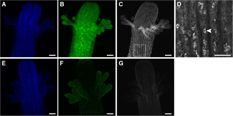
Fig. 5.
Immunolocalization of Eef1a-FLAG. a-d Eef1aFLAG-P2A-eGFP transgenic gastrozooids fixed, permeabilized, and stained with anti-FLAG primary antibodies and Alexa 647 conjugated secondary antibodies, followed by DAPI, and imaged on a laser scanning confocal microscope. (a, b) DAPI staining and eGFP expression in the transgenic gastrozooid, respectively. c Anti-FLAG staining in the transgenic gastrozooid, showing strong, diffuse signal throughout with sparse, intensely stained cells. d An inset of panel C showing intensely stained cells with polygon-like morphology (e.g., arrowhead). e-g Nontransgenic controls imaged and analyzed with identical settings. (e, f) DAPI staining and green fluorescence, respectively. Nematocyst capsules exhibit bright autofluorescence. g Anti-FLAG staining, showing faint background staining. Scale bars = 100 μm, except for (d), which is 50 μm

