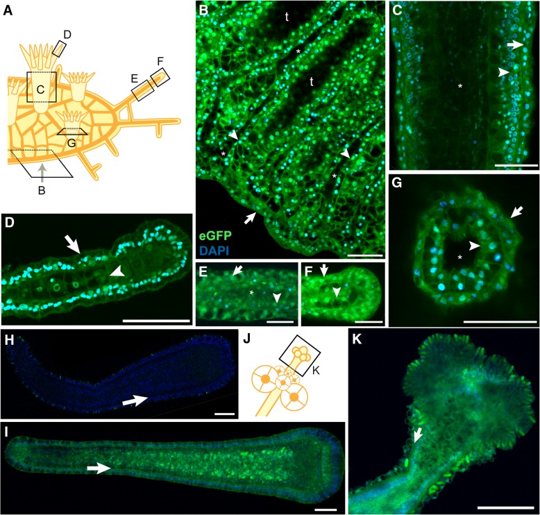
Fig. 6.
No evidence of variegated expression of the Eef1aFLAG-P2A-eGFP transgene in epithelial cell layers. a Schematic of colony showing orientation of multiphoton micrographs in b-g. b-g Multiphoton micrographs of Eef1aFLAG-P2A-eGFP expressing colonies stained with DAPI. Epidermal cell layers are indicated with arrows, while gastrodermal cell layers are indicated with arrowheads. b Edge of stolonal mat. Focal plane is through the center of the gastrovascular canals, leaving areas of the stolonal mat lacking canals dark because they are below the plane of focus (“t”). Asterisks denote lumens of gastrovascular canals. c Body of gastrozooid. Asterisk indicates lumen. d End of tentacle. e Middle of stolon. Asterisk denotes stolon lumen. The lumen has collapsed toward the left of the image. f Tip of stolon. g Base of polyp imaged through bottom of colony. Asterisk indicates lumen. h 120 hpf eGFP- larva from the 357 cross. Arrow denotes border of ectodermal and endodermal cell layers. i 120 hpf eGFP+ larva from the 357 population. Arrow same as in h. j Schematic of female gonozooid, showing plane of image in (k). k Confocal image of head of female gonozooid. Arrow denotes border of epidermal and gastrodermal cell layers. All scale bars = 50 μm

