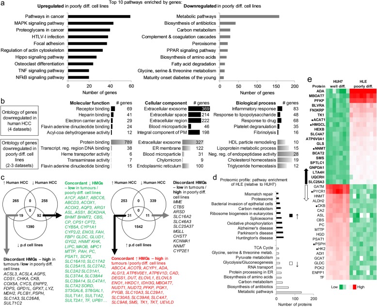
Fig. 2.
Functional annotation analysis, metabolic gene and proteomic profile of HCC cell lines. a Topmost pathway enrichment of genes upregulated (n = 1584) or lowly expressed (n = 1501) in poorly differentiated relative to well-differentiated HCC cell lines. P < 0.05 in 2–3 of the cell line datasets. Analysis was performed with DAVID v6.8, reference: Kyoto Encyclopedia of Genes and Genomes (KEGG). HTLV – Human T-lymphotropic virus, NFkB – nuclear factor kappa B, PPAR – peroxisome proliferator-activated receptor, TNF – tumor necrosis factor. b Topmost gene ontology (GO) for genes (n = 1547) consistently downregulated in all 4 human HCC microarrays datasets (GSE14520, GSE25097, GSE1898, GSE55092), compared to the GO profile of poorly differentiated cell lines. # - number of (genes). ER – Endoplasmic reticulum, HDL – high density lipoprotein, PM – plasma membrane, ‘Neg. – negative’ ‘Transcript.’ – transcriptional, ‘reg.’ – regulation. c Venn diagram showing human HCC tissue-derived metabolic genes (HMGs) expressed concordantly (i.e. mimicked) or discordantly by the poorly differentiated cell lines (2–3 datasets, P < 0.05). ↑ – upregulated; ↓ – downregulated. Note: the discordant genes are more mimicked by well-differentiated cell lines. A full list of the 92 concordant ↓ HMGs are contained in Additional file 6: Table S5. d Topmost pathway enrichment of proteins more expressed in HUH7 or HLE cells (FDR adjusted P < 0.05). e 20 most highly or lowly expressed HMGs-encoded proteins in the cell lines (FDR adjusted P < 0.05, mean fold difference > 2, HUH7 = 7, HLE = 8 samples). Arrows point to their encoding gene expression pattern in human HCC tissues. ● expressed in HCC in the opposite direction to the arrows

