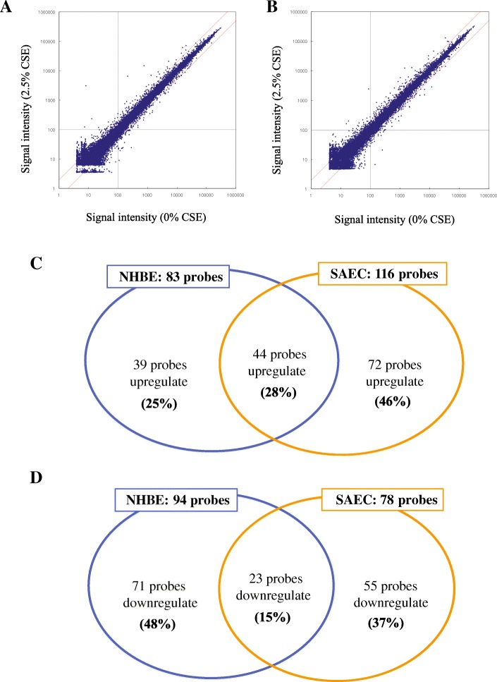
Fig. 1.
Gene-expression profiles after exposure to CSE in SAECs and NHBEs. Two different batches of SAECs and NHBEs were used. Cells were exposed to 2.5% CSE for 24 h or not (controls). Scatter plot of (a) NHBEs and (b) SAECs. The vertical axis represents the relative signal intensity in CSE-exposed cells, and the horizontal axis represents the control (without CSE exposure). Both axes show log scales. c Venn diagram of probes showing significant upregulation in SAECs and NHBEs after CSE exposure. d Venn diagram of probes showing significant downregulation in SAECs and NHBEs after CSE exposure. CSE, cigarette smoke extract; SAECs, small airway epithelial cells; NHBEs, normal human bronchial epithelial cells.

